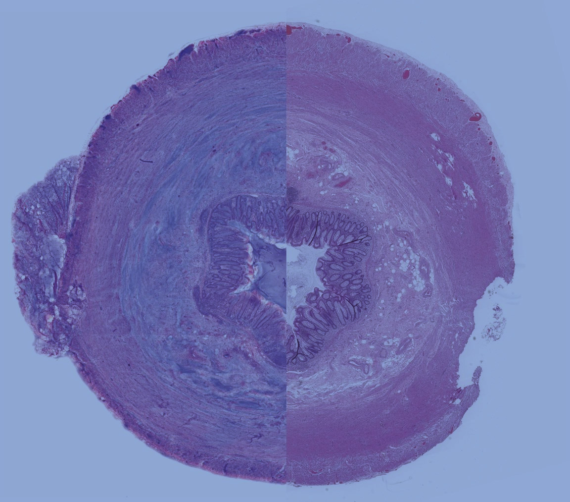
Histolix has developed a novel point-of-care histopathology solution enabling direct to digital diagnosis of tissue in surgery suites and at the clinical point-of-care, delivering high quality digitized images to permit rapid diagnosis. This can eliminate the need for time consuming and expensive histologic procedure required to produce paraffin sections on glass slides. It could enable same-day patient diagnosis and potentially reduce the requirement of additional surgical procedures, providing direct-to-digital image, slide free pathology designed to be created within five minutes of tissue presentation versus the traditional eight hours to days of required processing for whole slide imaging.


Time to Diagnosis
Led by longtime healthcare industry executives with multiple commercial exits in coordination with on ongoing, pioneering research by well-known University of California, Davis pathology researchers, Histolix provides the disruption needed for the growth of digital pathology across multiple use cases. With millions of grant and private investment dollars already deployed, Histolix technology is providing rapid, cost-effective, non-destructive intrinsically digital, slide-free tissue histology without compromising the quality of downstream molecular testing.


Histolix patented “direct read” technology provides a new intrinsically digital, slide-free histopathology solution that can be deployable in pathology labs, bypassing current workflow issues and potentially eliminating up to 30% of unnecessary repeated surgical procedures. To prove the breadth of this direct read value proposition, Histolix is undertaking a 100-tissue sample equivalency validation study with over 20 different tissue types. We expect to prove equal to or improved diagnostic quality with immediate read vs. the normal days of tissue processing, potentially eliminating the need for slide-based imaging completely.

